0
$\begingroup$

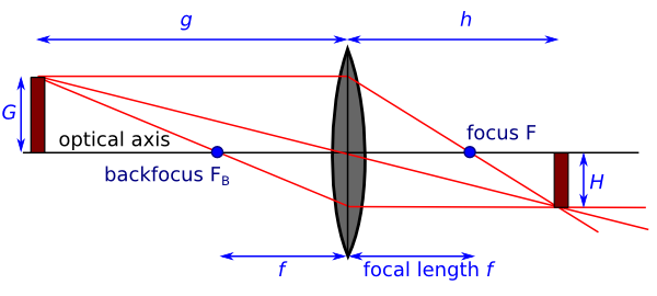
It is well known how an image is produced in general: You need at least two rays (in geometric optics) to form it, see the image next:

img2

But I wonder about these images:

img

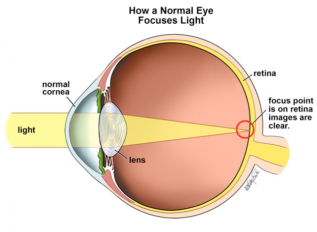
As you can see, in the upper picture, the generated image on the retina is achieved by, let's say, two rays that meet in a focus around the center of the eye. But in the lower picture, the image is generated while the focus is on the retina, so there are, in terms of geometric optics, no two rays to construct an image. It seems, that the lower version results in clearer images. From my understanding, there shouldn't be an image.

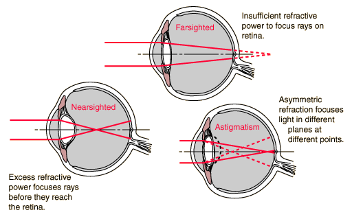
Using more sources provide a more insight view in regard to medical aspects:

img3

So the mechanism in picture 1 seems to be an error of the eye. However, according to common geometric optics, there are two rays needed that do not meet in the focus:

img4

So where am I'm wrong? I know it's quite basics but I can't get my head around it. Maybe it's more a question of medical matters as rays, focused on the retina, will deliver images behind it? Or is it physics I lack?

$\endgroup$
5
  • 1
    $\begingroup$ In geometric optics you can use two rays provided you have the focal point (and optic axis). I think all these images here are are for illustrative purposes only. Note that the last diagram has what you need to trace the image location (and only two rays are needed there). $\endgroup$
    – joseph h
    Commented Jun 29, 2021 at 8:37
  • $\begingroup$ but why do I have a clear image, when the focus point is on the retina, like in picture 2? $\endgroup$
    – Ben
    Commented Jun 29, 2021 at 8:53
  • 1
    $\begingroup$ Because that is what “in focus” means. Ie., when the image comes together on the retina. In the middle images they are not, so they are not in focus. $\endgroup$
    – joseph h
    Commented Jun 29, 2021 at 8:57
  • $\begingroup$ Well, does this mean, e.g., that, in the case of the sketch of the geometrical optics, one has an unclear image? $\endgroup$
    – Ben
    Commented Jun 29, 2021 at 9:18
  • $\begingroup$ See the answer below by Gandalf. Cheers. $\endgroup$
    – joseph h
    Commented Jun 29, 2021 at 9:26

1 Answer 1

1
$\begingroup$

Diagram 1 shows how a normal eye focuses diverging rays from a near object onto the retina to form an inverted image. Just two rays (1 and 2) are shown from the top of the tree and two rays (3 and 4) from the bottom, but many more rays could have been shown - the normal eye will focus all rays that come from a common point on the object onto a single image point on the retina.

Diagram 2 shows how a normal relaxed eye focuses parallel rays from a point at infinity onto a single point on the retina. Again, just two rays are shown for illustration, but more rays could have been shown. Note how the rays in Diagram 2 are bent less than the rays in Diagram 1, and the lens of the eye has a different shape.

To focus on a near object, the ciliary muscles around the lens contract, which decreases the tension on the lens and allows it to become rounder, with a shorter focal length. This process is called accommodation. Accommodation allows the normal eye to focus rays from objects at a wide range of distances - from infinity down to less than 10 cm - into an image that falls on the retina. Note that the lens should actually be rounder in Diagram 1 and flatter in Diagram 2 - the shape of the lens in the diagrams is incorrect.

The third set of diagrams shows how the lens in the relaxed eye can have a focal length that is too short (short-sightedness or myopia) or too long (far-sightedness or hyperopia). Short-sighted people have clear vision for near objects and blurred vision for far objects; far-sighted people have the opposite. The third case, astigmatism, is when the lens does not bring rays from a common point to a single focus at all, resulting in blurred vision at any distance.

$\endgroup$
3
  • $\begingroup$ Thank you! Why will there be an image in the case of diagram 2? In terms of geometric optics, just simply, because two light rays meet? $\endgroup$
    – Ben
    Commented Jun 29, 2021 at 10:31
  • 1
    $\begingroup$ @Ben Diagram 2 shows rays coming from one point on a distant object (this is why the rays are shown as parallel) and they are focused to one point on the retina. Rays coming from another point on the distant object will come into the lens at a different angle, so will be focussed to a different point on the retina - but this isn't shown in the diagram. $\endgroup$
    – gandalf61
    Commented Jun 29, 2021 at 10:38
  • $\begingroup$ ah, got it. thanks! $\endgroup$
    – Ben
    Commented Jun 29, 2021 at 11:00

Your Answer

By clicking “Post Your Answer”, you agree to our terms of service and acknowledge you have read our privacy policy.

Not the answer you're looking for? Browse other questions tagged or ask your own question.