In my efforts to characterise a 0.67 NA microscope objective (working distance of about 15mm, effective focal length of 25mm), I have placed a 20 micron precision pinhole at the focal plane of the objective, and back illuminated the pinhole with a 767nm laser. The light coming out of the objective is focused onto the imaging plane (camera plane) with a F=1 metre lens.
By my understanding, the image field $U_i$ should be a convolution of the Point Spread Function (PSF) $h$ and the object field $U_o$, i.e.
$$ U_i(x_i,y_i) \propto \int_{-\infty}^\infty\int_{-\infty}^\infty h(x_i-\xi, y_i-\eta) U_o(\xi,\eta)\, d\xi d\eta\, , $$
where the proportional sign is for good measure as there are some coefficients in front, but that does not affect the profile. The image intensity profile is given by
$$ I_i(x_i, y_i) = |U_i(x_i, y_i)|^2\,. $$
In my case the microscope objective has a circular aperture, and that means that the PSF $h$ is given by the airy function, i.e.,
$$ h(x,y) = \frac{J_1 \left( \frac{2\pi}{\lambda} NA \sqrt{x^2 + y^2}\right)}{\sqrt{x^2 + y^2}} \, , $$
where $J_1$ is the Bessel function of the first kind, order one.
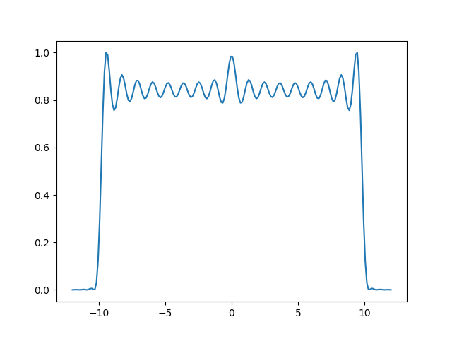
I have plotted out the intensity profile (gauss quad integral) that I expect for the current pinhole and wavelength, taking into account the magnification of the system, and I got the following plot.
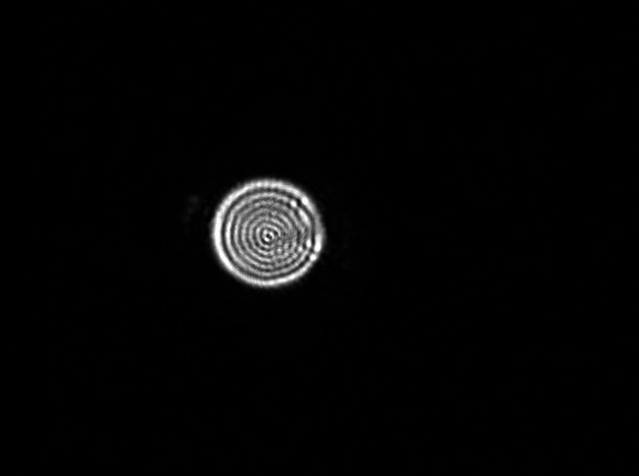
However, this differs from my measurements taken in experiment:
The size of the image is approximately 20 microns (pixel size = 3.75/40 microns). The number of peaks somewhat represent what is shown from the plot above. What concerns me more is how the intensity profiles goes to zero between peaks, unlike what the theory predicted.
Is my understanding of the current theory incorrect?


